Applications of QUANTIFOIL® Holey Carbon Films | ||
In electron microscopy, support foil with a high percentage of open area is used in order to minimize the total specimen thickness or to eliminate the object distortions arising from interactions with a support. Thus, perforated foil is used to bear an ultra-thin carbon support foil or to directly support a large object, such as a powder particle, in the case of electron diffraction and energy loss spectroscopy, and electron spectroscopic and dark field imaging. It is also used when small objects, such as biomacromolecular
complexes, are studied in a free hanging layer of sustaining medium. This
medium is vitreous ice in cryo-EM. | ||
Preparation of vitrified specimensSpecial measures may be necessary in order to obtain an optimally thin ice film that is of constant thickness over a hole. The distribution of the aqueous solution over a grid, that over a hole, and the thickness of film within a hole will depend on, among other factors, the surface properties of the foil, i.e. its wetting properties, and on the relative humidity of the environment after blotting and before freezing. Untreated QUANTIFOIL® tends to be hydrophobic. The degree of hydrophilicity can be influenced by glow-discharging in residual air, at which parameters such as the duration of glow-discharging and the time elapsed before specimen preparation, can be varied. Some cryo-microscopist find the wetting properties of holey film best when it is metal-coated. The relative humidity around the specimen can be influenced by a flow of humid air or with an environmental chamber. A relative humidity of 100% can be attained with the environmental chamber of the Vitrobot (Maastricht Instruments BV, The Netherlands). The Vitrobot is a fully PC-controlled device for vitrification of aqueous samples. The vitrification robot yields optimally thin ice films with a constant thickness over a hole and consequently with a homogeneous particle distribution. Just as QUANTIFOIL® facilitates high throughput of image data by automated acquisition - see below -, the Vitrobot enables high throughput of samples. |
Low-dose Electron Microscopy |
|
QUANTIFOIL® greatly simplifies the carrying out of low-dose procedures. The location of the area(s) used for focussing, relative to the area that is recorded, need(s) to be set only once for a specimen. | |
| |
|
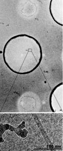
Vitrified specimen of
tobacco mosaic virus (TMV) prepared using QUANTIFOIL® holey carbon
film. |
Reprinted with permission from Elsevier Science, Ultramicroscopy 74, 75-81, Ermantraut, Wohlfart, Tichelaar, Perforated support foils with pre-defined hole size, shape and arrangement (1998), copyright 1998 |
| ||
Automated image acquisition | ||
QUANTIFOIL®
allows image acquisition to be performed automatically. |
|
|
|
Courtesy of Dr. B. Carragher |
|
The procedure includes |
| |
|
(1) |
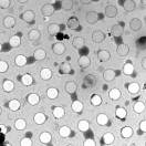
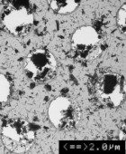
Acquisition of a low magnification image (left image, showing TMV embedded in vitreous ice suspended over 2 µm holes) |
|
|
(2) |
Automatic identification of holes containing ice of the suitable thickness (note the cross in the left image) |
|
|
(3) |
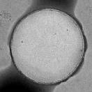
Acquisition of an intermediate magnification image of an identified hole (middle) |
|
|
(4) |
Identification of features of interest within the hole |
|
|
(5) |
Focussing at the magnification to be used for the picture |
|
|
(6) |
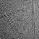
Acquisition of a high magnification image (right, showing microtubules in this case) |
|
| ||
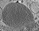
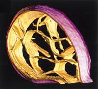
Cryo-electron tomographic reconstructionA reconstruction of Neurospora mitochondria was carried out by
Nicastro and collegues (Max-Planck-Institute for Biochemistry,
Martinsried). | ||
|
An electron micrograph of a zero-degree projection of a tilt series; the edge of the hole in QUANTIFOIL® is seen at the bottom. |
|
| ||
|
A central x-y slice through the reconstructed volume. |
|
| ||
|
A view of the reconstruction, showing that the crista membrane that forms a three-dimensional network of interconnected lamellae is constantly bound with the inner boundary membrane (both shown in yellow). The outer membrane (magenta) is separated from the inner one by a narrow band of remarkably constant width. Courtesy of Dr. D. Typke Reprinted with permission of Academic Press, J. Struct. Biol. 129,48-56, D. Nicastro et al., Cryo-electron tomography of Neurospora mitochondria (2000), copyright 2000 | |
| ||
|
First application of cryo-EM to cement | ||||
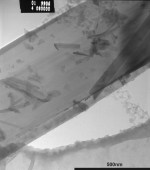
At the F. A. Finger Institute for Building Materials Science (Bauhaus University Weimar), hitherto unknown structural properties of calcium silicate hydrate phases (C-S-H phases) were discovered by freezing freshly prepared EM specimens of cement and keeping in the frozen state during microscopy (Rößler et al., in preparation). The C-S-H phases account to the strength in ordinary Portland cement and concrete. Frozen-hydrated specimens of C-S-H phases on
QUANTIFOIL®: | ||||
| ||||
| ||||
|
|
| |
Low-energy electron point source - LEEPS- microscopy | ||
In low-energy electron point source microscopy a perforated
foil with a regular pattern is required in order to be able to distinguish
an object which is spanned over a hole. An object cannot be discriminated
from the support in the case of conventional holey film. H.-W.
Fink & C. Schönenberger (University of Basel) used
QUANTIFOIL® for the
measurement of electrical current through DNA
molecules. | ||
|
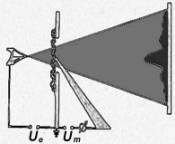
A schematic representation of a LEEPS microscope with a manipulation tip between specimen and detector. |
|
| ||
|
A projection image of ropes of lambda DNA spanning a 2 µm hole. Courtesy of Dr. H.-W. Fink Reprinted with permission from Nature, Nature 398, 407-410, H.-W. Fink & C. Schönenberger, Electrical conduction through DNA molecules (1999), copyright 1999 Macmillan Magazines Ltd. |
|
| ||
| ||
LiteratureCarragher B, Kisseberth N, Kriegman D, et
al. Ermantraut E, Wohlfart K, Tichelaar W Fink H-W, Schönenberger C Gao H, Spahn CMT, Grassucci RA, Frank J Glaeser RM Ledoux G, Amans D, Gong J, et al. Nicastro D, Frangakis AS, Typke D, et
al. Pulokas J, Green C, Kisseberth N, et al. Reviakinc I, Bergsma-Schutter W, Morozov AN, et
al. Rouiller I, DeLaBarre B, May AP et
al. Tao Y, Zhang W Unger VM van Heel M, Gowen B, Matadeen R, et
al. Volkmann N, Amann KJ, Stoilova-McPhie S, et
al. Weierstall U, Spence JCH, Stevens M, et
al. | ||
|